


POSTER - ECDP 2022
A tool for automatic detection of mitoses in breast cancer

By: Clara Simmat (Primaa), Stéphane Sockeel (Primaa), , Marie Sockeel (Primaa), Nicolas Pozin (Primaa), Éloi Bréhin (Primaa), Loris Guichard (Hôpital Bicêtre), Sophie Prévot (Hôpital Bicêtre), Magali Lacroix-Triki (Institut Gustave Roussy), Catherine Miquel (Hôpital Saint-Louis).
Material & methods: from data collection to a detection pipeline
Train dataset
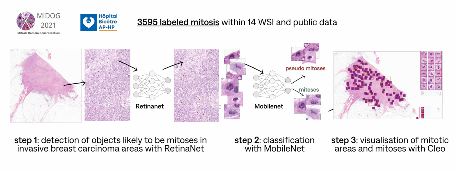
A two-step localisation model trained on in-house labeled data:
- a RetinaNet detector to detect mitotic candidates in invasive breast carcinoma areas,
- a MobileNet classifier to refine these detections.
Cleo, our visualisation tool, displays detected mitoses and areas of highest mitotic density (hotspots).
Algorithmic and clinical performances
Algorithmic performances to find hotspots
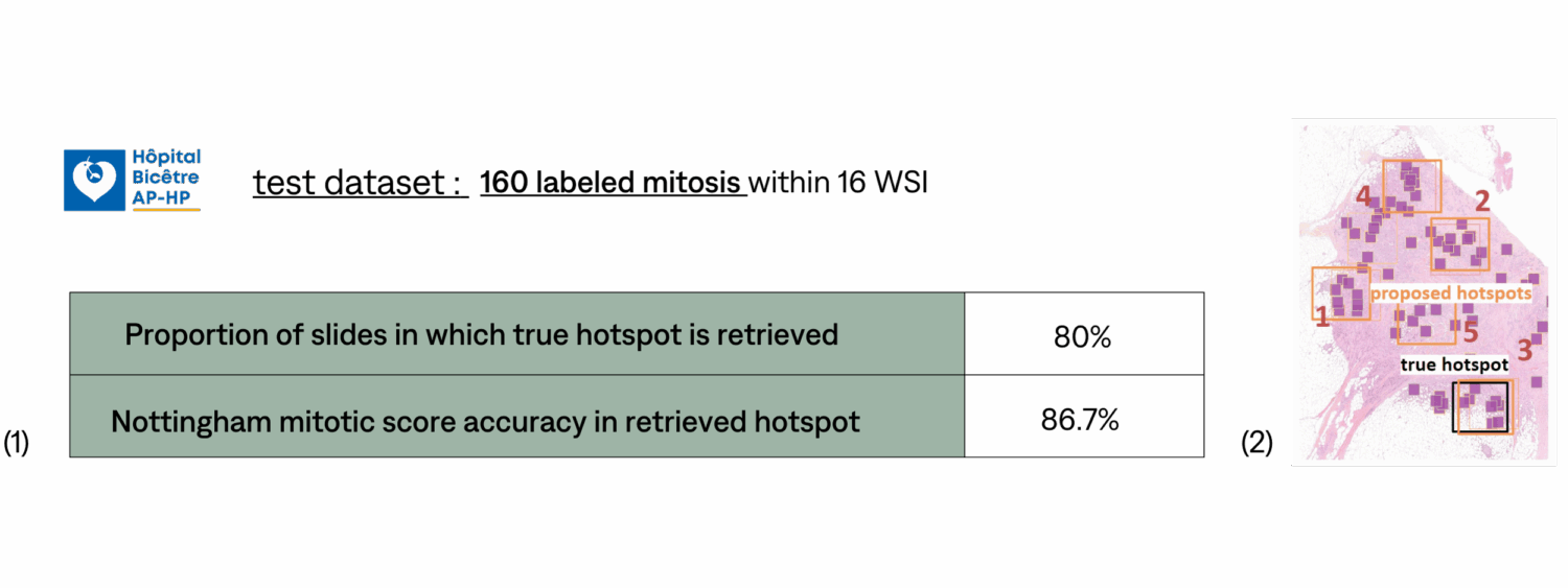
(1) The five 2mm² areas with highest mitotic density are retrieved by the algorithm and compared with ground-truth hotspot for each slide.
(2) Previous studies showed moderate interobserver agreement for the hotspot selection (kappa=0.53). Such discrepancy could be answered by our solution.
Clinical performances in a pre-selected hotspots
Study dataset : 856 labeled mitosis within 2mm² areas in 50 WSI.
For this clinical study, 4 pathologists labeled mitosis on 50 pre-selected 2mm² areas from 50 different slides, with and without AI-results displayed. The ground-truth was established with mitosis labeled without AI results displayed, but the inter and intra reproducibilities in mitotic detection make ground truth determination of mitotic figures a major challenge.
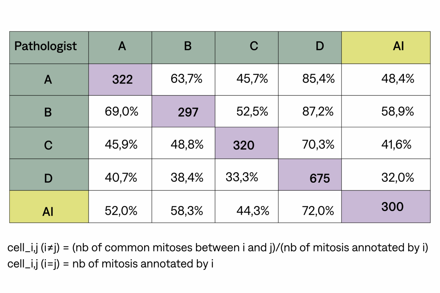
Reproductibility inter obs. (mitotic figures)
Among mitotic figures detected by AI, 44.3% to 72.0% are also found by pathologists. Among mitotic figures detected by pathologists, 32.0% to 58.9% are also found by AI. Similar metrics are computed for pathologists.
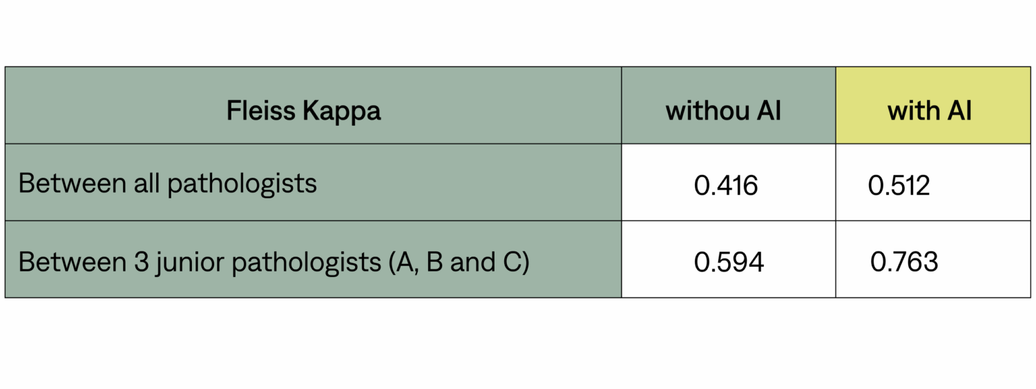
Reproductibility inter obs. (mitotic score)
Conclusion
Our automatic detection pipeline tool is good at picking relevant hotspots. Regarding detection of mitotic figures, tt has a comparable level of concordance than any other pathologist would. We also show that Fleiss Kappa on mitotic scores is improved when pathologists are assisted by AI. A clinical study to assess if our tool can help pathologists in determining the Notthingam mitotic score in routine is in progress.
References
Ibrahim A, Lashen A, Toss M, Mihai R, Rakha E. Assessment of mitotic activity in breast cancer: revisited in the digital pathology era. J Clin Pathol (2022) — Elston EW, Ellis IO Method for grading breast cancer.Journal of Clinical Pathology (1993) — Ibrahim A., Lashen A.G., Katayama A. et al. Defining the area of mitoses counting in invasive breast cancer using whole slide image. Mod Pathol (2021).