


POSTER - ECP 2022
Automatic detection of microcalcifications in Whole Slide Images

By: Marceau Clavel (Primaa), Nicolas Pozin (Primaa), Stephane Sockeel (Primaa), Marie Sockeel (Primaa), Catherine Miquel (APHP France).
Data
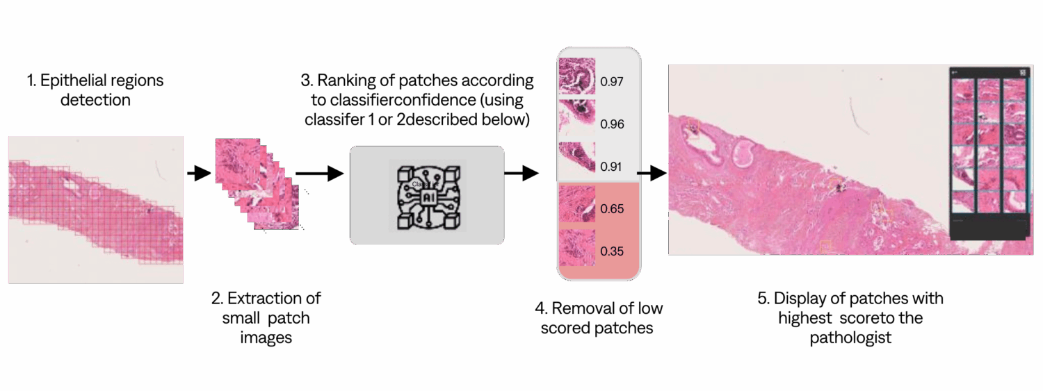
Microcalcifications are calcium deposits in breast products. If the mammography reveals microcalcifications, the anatomopathologist should identify them. To help the pathologist, we built a microcalcification detection pipeline on Whole Slide Images. We compare two patch-based classification methods against a set of custom metrics designed to measure the help brought to pathologists.
We collected 1615 training slides from Center A , among which 174 were containing calcifications. Our test data comes from 4 different medical centers:

Method
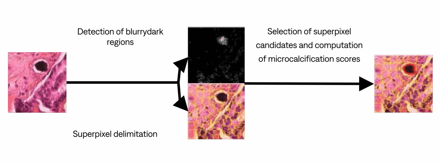
Classifier 1 (trained on 5 slides) Image processing method.
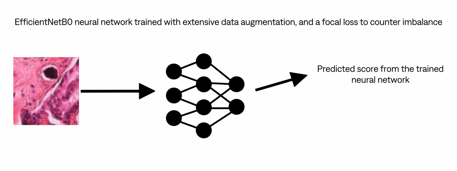
Classifier 2 (trained on 1615 slides) Deep Learning method.
Results
In the pathologist workflow, microcalcification detection is a binary problem: the pathologist should detect whether a biopsy contains microcalcifications or not. To assess the help brought to pathologist, we introduce the following metrics:
Microcal_in_top_16: 1 if the microcalcification is suggested in the 16 first patches. (higher is better)
FP_in_top_16: Number of false positive among the 16 first patches. (lower is better)
Rank_top: Rank of the first True Positive among the 16 first patches. (lower is better)

Discussion
Results show that Deep Learning classifier outperforms Image processing classifier. Deep Learning classifier has similar results on seen and unseen centers, demonstrating the generalization ability of this approach. Image processing classifier requires less training data, and results show that it can be a valid lightweight option. Depending on the availability of labeled data, performance needs and computing resources, both classifier can be considered.
Conclusion
The developed automatic pipeline can be integrated seamlessly in any digital pathologist workflow, helping them to spot microcalcifications efficiently on whole slide images. This pipeline can be applied to other small object detection with sufficient available data.